entellus Medical
3563-002 XprESS ENT Dilation System Instructions for Use
4 Pages
Preview
Page 1
3563-002
XprESS™ ENT Dilation System
INSTRUCTIONS FOR USE ALL INSTRUCTIONS, PRECAUTIONS AND WARNINGS SHOULD BE CAREFULLY READ AND UNDERSTOOD BEFORE USE. FAILURE TO DO SO MAY RESULT IN COMPLICATIONS.
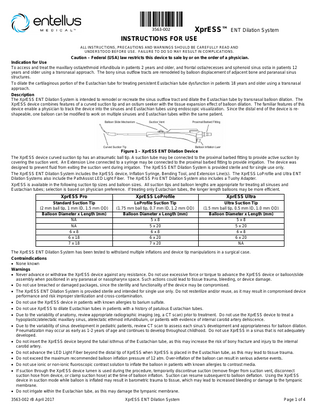
Caution – Federal (USA) law restricts this device to sale by or on the order of a physician. Indication for Use To access and treat the maxillary ostia/ethmoid infundibula in patients 2 years and older, and frontal ostia/recesses and sphenoid sinus ostia in patients 12 years and older using a transnasal approach. The bony sinus outflow tracts are remodeled by balloon displacement of adjacent bone and paranasal sinus structures. To dilate the cartilaginous portion of the Eustachian tube for treating persistent Eustachian tube dysfunction in patients 18 years and older using a transnasal approach. Description The XprESS ENT Dilation System is intended to remodel or recreate the sinus outflow tract and dilate the Eustachian tube by transnasal balloon dilation. The XprESS device combines features of a curved suction tip and an ostium seeker with the tissue expansion effect of balloon dilation. The familiar features of this device enable a physician to track the device into the sinuses and Eustachian tubes using endoscopic visualization. Since the distal end of the device is reshapeable, one balloon can be modified to work on multiple sinuses and Eustachian tubes within the same patient.
Figure 1 – XprESS ENT Dilation Device The XprESS device curved suction tip has an atraumatic ball tip. A suction tube may be connected to the proximal barbed fitting to provide active suction by covering the suction vent. An Extension Line connected to a syringe may be connected to the proximal barbed fitting to provide irrigation. The device was designed to prevent fluid from exiting the suction vent during irrigation. The XprESS ENT Dilation System is provided sterile and for single use only. The XprESS ENT Dilation System includes the XprESS device, Inflation Syringe, Bending Tool, and Extension Line(s). The XprESS LoProfile and Ultra ENT Dilation Systems also include the PathAssist LED Light Fiber. The XprESS Pro ENT Dilation System also includes a Tuohy Adapter. XprESS is available in the following suction tip sizes and balloon sizes. All suction tips and balloon lengths are appropriate for treating all sinuses and Eustachian tubes; selection is based on physician preference. If treating only Eustachian tubes, the longer length balloons may be more efficient.
XprESS Pro
XprESS LoProfile
XprESS Ultra
Standard Suction Tip (2 mm ball tip, 1 mm ID, 1.5 mm OD) Balloon Diameter x Length (mm) NA NA 6x8 6 x 18 7 x 18
LoProfile Suction Tip (1.75 mm ball tip, 0.7 mm ID, 1.2 mm OD) Balloon Diameter x Length (mm) 5x8 5 x 20 6x8 6 x 20 7 x 20
Ultra Suction Tip (1.5 mm ball tip, 0.5 mm ID, 1.0 mm OD) Balloon Diameter x Length (mm) 5x8 5 x 20 6x8 6 x 20 NA
The XprESS ENT Dilation System has been tested to withstand multiple inflations and device tip manipulations in a surgical case. Contraindications • None known Warnings • Never advance or withdraw the XprESS device against any resistance. Do not use excessive force or torque to advance the XprESS device or balloon/slide assembly when positioned in any paranasal or nasopharynx space. Such actions could lead to tissue trauma, bleeding, or device damage. • Do not use breached or damaged packages, since the sterility and functionality of the device may be compromised. • The XprESS ENT Dilation System is provided sterile and intended for single use only. Do not resterilize and/or reuse, as it may result in compromised device performance and risk improper sterilization and cross-contamination. • Do not use the XprESS device in patients with known allergies to barium sulfate. • Do not use XprESS to dilate Eustachian tubes in patients with a history of patulous Eustachian tubes. • Due to the variability of anatomy, review appropriate radiographic imaging (eg, a CT scan) prior to treatment. Do not use the XprESS device to treat a hypoplastic/atelectatic maxillary sinus, atelectatic ethmoid infundibulum, or patients with evidence of internal carotid artery dehiscence. • Due to the variability of sinus development in pediatric patients, review CT scan to assess each sinus’s development and appropriateness for balloon dilation. Pneumatizaton may occur as early as 1-2 years of age and continues to develop throughout childhood. Do not use XprESS in a sinus that is not adequately developed. • Do not insert the XprESS device beyond the tubal isthmus of the Eustachian tube, as this may increase the risk of bony fracture and injury to the internal carotid artery. • Do not advance the LED Light Fiber beyond the distal tip of XprESS when XprESS is placed in the Eustachian tube, as this may lead to tissue trauma. • Do not exceed the maximum recommended balloon inflation pressure of 12 atm. Over-inflation of the balloon can result in serious adverse events. • Do not use ionic or non-ionic fluoroscopic contrast solution to inflate the balloon in patients with known allergies to contrast media. • If suction through the XprESS device lumen is used during the procedure, temporarily discontinue suction (remove finger from suction vent, disconnect suction hose from device, or clamp suction hose) at the time of balloon inflation. Suction can resume subsequent to balloon deflation. Using the XprESS device in suction mode while balloon is inflated may result in barometric trauma to tissue, which may lead to increased bleeding or damage to the tympanic membrane. • Do not irrigate within the Eustachian tube, as this may damage the tympanic membrane. 3563-002 rB April 2017
XprESS ENT Dilation System
Page 1 of 4
• As in any upper airway procedure or sinus surgery, do not have patient use CPAP until the physician has confirmed that the tissue is adequately healed. CPAP use prior to soft tissue healing may result in facial and/or neck swelling due to subcutaneous emphysema. • Do not clean the XprESS device with anti-microbial agents as the compatibility of the XprESS device with these agents has not been tested. • The XprESS device has been tested only with the Fiagon Navigation System. Do not attach the XprESS device to other image guidance systems, as use with other systems may result in inaccurate device positioning. Refer to System Operation 1.b for instructions on how to connect XprESS to the Fiagon system. • The XprESS device has been tested only with the Entellus Inflation Syringe. Do not use other inflation devices with the XprESS device, as doing so may result in serious patient injury. Precautions o Store the XprESS device components in a cool and dry place. Never use a device that is beyond its expiration date. o Handle the XprESS device with care. Prior to use, and during the procedure, inspect the packaging and components for bends, kinks, or other damage. Discontinue the use of the XprESS device if it may have been damaged. o Select a balloon diameter that will result in expansion of the tissue post dilation. Do not select a balloon diameter that is larger than the bony margins of the outflow tract as this may damage the balloon. o Pay special attention when advancing or withdrawing the balloon and slide assembly. If resistance is encountered, use endoscopy or direct visualization to help guide device out of the paranasal or nasopharynx space and then attempt to alleviate the resistance. If the cause of resistance cannot be determined, do not use the XprESS device. o Use direct endoscope visualization with or without PathAssist LED Light Fiber or Light Fiber to ensure accurate placement of the balloon prior to dilation. If balloon location cannot be verified, image guidance or fluoroscopy can be used. If balloon location still cannot be verified, the balloon should not be inflated. o Consider using self-limiting radiation exposure equipment when employing fluoroscopy to confirm device placement. Ensure the equipment is calibrated and maintained according to the equipment manufacturer’s user manual. o Use techniques for reducing fluoroscopic exposure when using fluoroscopy. Examples are applying pulsed beam settings, increasing target-to-panel distance, using posterior-anterior projection, and using appropriate lead shield protection. Total fluoroscopy time should be limited to 30 minutes. o When fluoroscopy is used, especially in children, minimize radiation dose to the lens of the eye and other proliferating tissues due to the potential for cataract formation or injury to the surrounding tissue. o Do not advance or withdraw a guidewire through the XprESS Pro or LoProfile suction/irrigation lumen against resistance. This could lead to device damage. o Be aware that guidewires (including Fiagon GuideWires) do not track through the XprESS Pro or LoProfile when they are bent in the recommended maxillary configuration or through the XprESS Ultra in any configuration. Other methods can be used to obtain confirmation of the treatment area, such as use of the PathAssist Light Fiber, direct visualization of the XprESS device with an aid of an endoscope, or fluoroscopic imaging of the XprESS tip. o Use standard larger suction tubes for removal of thick secretions or other materials. XprESS Pro has a 1 mm ID comparable to that of a 5F suction tube. XprESS LoProfile has a 0.7 mm ID comparable to that of a 4F suction tube. XprESS Ultra has a 0.5 mm ID comparable to that of a 2.5F suction tube. All are capable of removing blood and thin mucous. o Fully deflate the balloon and retract the balloon slide assembly before withdrawing the XprESS device from the paranasal or nasopharynx space. o Use only liquid contrast or saline solution for inflation. Do not inflate with air. o Consider using a new balloon if cross-contamination between sinuses or Eustachian tubes is a concern. Adverse Effects Possible adverse effects include, but are not limited to, the following: • Complication from anesthesia • Cavernous sinus syndrome • Revision surgery • Damage to the lamina papyracea • Tinnitus • Damage to the lacrimal sac affecting tearing • Damage of the orbital wall or other structures of • Pneumocephalus • Damage to the Eustachian tube the eye • Patulous Eustachian tube • Bruising and swelling • Cerebrospinal fluid leak • Tissue inflammation • Permanent hearing loss • Loss of vision or diplopia (double vision) • Fever and infection • Carotid artery damage • Pain • Continued or worsening symptoms • Tympanic membrane damage • Bleeding Supplies The following supplies are not provided with the XprESS ENT Dilation System and should be available and prepped prior to use of the device. − Appropriate endoscopes and compatible camera system − ≥50 mL of sterile saline solution, sterile fluoroscopic contrast solution, or sterile water − Needles and syringes as required for injections − 20-30 mL syringe and Extension Line (if irrigation is to be performed) − Suction system − Other supplies or medication as established by laboratory protocol − If the use of a sterile guidewire is desired (compatible with the XprESS Pro), the recommended guidewire should be sterile and ≤0.035 inches in diameter with a minimum length of 50 cm. Example of a guidewire that meets these requirements is the Entellus Medical Sinus Guidewire. − If desired, Entellus Medical PathAssist™ LED Light Fiber, Light Fiber™, or Light Seeker Optional Equipment − Fluoroscopy may be used in conjunction with the endoscope if desired. − Fiagon Navigation System and GuideWires (GuideWire and GuideWire − Refer to appropriate Instructions for Use and safety procedures when 0.6 are compatible with XprESS Pro; GuideWire 0.6 is compatible with preparing and using equipment. XprESS LoProfile) Instructions for Use System Preparation 1. Prepare the Inflation Syringe and Extension Line a. Remove the Inflation Syringe and Extension Line from its sterile package. Note the 3 referenced Inflation Syringe plunger positions:
Figure 2 - Plunger all the way in 3563-002 rB April 2017
Figure 3 - First Click position XprESS ENT Dilation System
Figure 4 -Second Click position (all the way out) Page 2 of 4
b. Begin with the Inflation Syringe plunger all the way in (Figure 2). c. Then submerge tip in sterile saline solution.
d. Fill Inflation Syringe by slowly drawing plunger back to second click position (all the way out) (Figure 4).
e. Attach an Extension Line to the filled Inflation Syringe.
f. Point the syringe tip towards the ceiling. Tap the Inflation Syringe until a large bubble is visible beneath the orange piston.
g. While still pointing the syringe tip towards the ceiling, push the plunger all the way in (Figure 2), to purge all air and fluid from the syringe.
h. Submerge the free end of the Extension Line in sterile saline solution. Slowly draw plunger back to the first click position (Figure 3) to fill the syringe.
2. Prepare XprESS ENT Dilation System. a. Remove the XprESS device from its sterile package. b. Remove and discard the balloon protector. c. Connect the free end of the prepped Extension Line to the XprESS balloon inflation luer. Note: Inspect the syringe barrel to ensure there is minimal air in the system. If excessive air remains in the system, repeat prepping process. Figure 5: Alignment between Distal Seal and Distal Mark d. Perform a test inflation of the system by depressing the plunger rod until the distal black seal on the orange piston is aligned with the distal black mark of the Inflation Syringe (See Figure 5). If the seal and black mark do not align, disconnect the Inflation Syringe and Extension Line and repeat the prepping process.
Alignment between the Distal Seal and the Distal Mark Corresponds to 12atm
e. Pull the plunger rod back to the 2nd click to apply a vacuum to the balloon. Ensure there is no air introduced into the system during deflation of the balloon. If a leak is detected and the source cannot be identified and corrected, do not use the XprESS device, Extension Line, and Inflation Syringe. Use new devices to complete the procedure.
Distal Seal
Distal Mark
f. If suction or irrigation is planned, connect the Extension Line to the proximal barbed fitting to add a flexible connector for suction or irrigation. Reshaping the XprESS Device Suction Tip to Treat Multiple Spaces
Orange Piston
− When treating multiple spaces, it is recommended to complete balloon dilation of the frontal or sphenoid sinuses or Eustachian tubes prior to treatment of the maxillary sinuses. − Frontal Sinuses: When treating the frontal recesses, a large radius curve similar to a frontal sinus seeker (Figure 6) is recommended. This is the shape/curve provided in the package. − Sphenoid Sinuses: When treating the sphenoid sinus ostia, a slight bend (Figure 7) is recommended. − Eustachian Tubes: When treating the Eustachian tubes, a bend of approximately 45° at the 2 cm mark (Figure 8) is recommended. − Maxillary Sinuses: When treating the maxillary ostia/ethmoid infundibula, a bend of approximately 120 - 135° (Figure 9) is recommended to gain access to the natural maxillary ostium. Use the included Bending Tool to achieve this geometry.
Figure 6: Frontal Bend Figure 7: Sphenoid Bend Figure 8: Eustachian Tube Bend − Small adjustments to the above bends may be considered to accommodate different patient anatomy. Using Bending Tool − The Bending Tool should be used to achieve the proper maxillary bend. The tool also provides a frontal and sphenoid bend configuration if needed. − Maxillary Bending with Bending Tool: Before shaping the maxillary bend, the device should be close to straight as shown for a Sphenoid Bend. With the Bending Tool in one hand, position the ball tip into the ball holder in the bending tool (Figure 10). Place a finger at about the 2 cm mark on the suction tip and use this finger to form the Maxillary Bend (Figure 11). Figure 10 – Start Maxillary Bend Patient Preparation
Figure 9: Maxillary Bend
Figure 11 – Finish Maxillary Bend
1. Patient preparation should be consistent with standard practice. 2. Anesthesia should be administered appropriately to allow patient tolerance. System Operation 1. Locate the sinus structure or Eustachian tube orifice using one of the following confirmation methods: a. Direct Visualization with or without Light Confirmation: Locate the treatment area using XprESS with or without LED Light Fiber, Light Fiber, Light Seeker, a standard sinus ostium seeker, and/or guidewire with the aid of an endoscope. Observe the location of the treatment area relative to the anatomical landmarks through the endoscope. Remove the Light Seeker, sinus ostium seeker, or guidewire after locating treatment area. Note: If using the PathAssist LED Light Fiber or Light Fiber, refer to the Instructions for Use (IFU) for complete instructions. b. CT Image Guidance: If further confirmation of the treatment area location is desired, CT image guidance using the Fiagon Navigation System and GuideWire or GuideWire 0.6 with XprESS Pro may be used. The Fiagon Navigation System and GuideWire 0.6 with XprESS LoProfile may also be used. i. If using the GuideWire with XprESS Pro, attach the Tuohy Adapter to the XprESS proximal barbed fitting. ii. Load the Fiagon GuideWire through the Tuohy Adapter and working lumen of XprESS until the tip of GuideWire aligns with the tip of XprESS. iii. Secure the GuideWire in place by tightening the Tuohy Adapter. iv. If using GuideWire 0.6 with XprESS Pro or LoProfile, load the GuideWire 0.6 through the working lumen of XprESS until the luer lock connector meets the proximal barbed fitting of XprESS. 3563-002 rB April 2017
XprESS ENT Dilation System
Page 3 of 4
v. Secure the luer lock connector on the proximal barbed fitting. vi. Refer to Fiagon Navigation System Instructions for Use. Note: Neither of the Fiagon GuideWires should be used with any XprESS device in the maxillary bend configuration. Note: Do not attach the XprESS device to other image guidance systems. c. Fluoroscopy: If further confirmation of the treatment area is desired, fluoroscopy may be used. Take two orthogonal views (AP and lateral). The XprESS device suction tip is stainless steel and is visible under fluoroscopy. The balloon will be proximal to the tip of the device. 2. Under endoscopic visualization, track the XprESS device to the same treatment area identified above. a. Position XprESS suction tip within the sinus ostia or within the cartilaginous portion of the Eustachian tube. Notes: Reference marks are located 1 and 2 cm from the tip of the device. The XprESS suction tip may be reshaped to aid in device positioning. Use device as a suction tool to maintain a clear visual field during device positioning. Cover suction vent with finger to allow suction. 3. Advance the balloon by fully advancing the balloon slide mechanism forward to position the balloon within the sinus opening or Eustachian tube. 4. Prior to inflating balloon, discontinue the use of suction (remove finger from suction vent, disconnect suction hose from device, or clamp suction hose) to decrease the risk of barotrauma. 5. Balloon dilation of the treatment site: a. Slowly depress the Inflation Syringe plunger rod to inflate the balloon. The pressure should be increased slowly (3-5 seconds) until the orange piston bottoms out (distal black seal of the piston reaches the distal black mark on the Inflation Syringe – see Figure 5). If these do not align, deflate the balloon and remove the XprESS device and perform a test inflation (as described in steps 2.d and 2.e of the System Preparation section). Alignment of the distal mark and distal seal will ensure that 12 atm of pressure is reached. Note: Do not use air or any gaseous medium to inflate the balloon. b. Inflate the balloon until the desired result is achieved or until it reaches 12 atm. Sinus Dilation: Inflate the balloon for up to 20 seconds (less than or equal to 20 seconds); observe that the balloon is inflated endoscopically. Eustachian Tube Dilation: Inflate the balloon for approximately 2 minutes by holding in the plunger rod; observe that the balloon is inflated endoscopically. Note: Do not exceed 12 atm. Warning: To avoid barometric trauma to tissue, do not use device in suction mode (remove finger from suction vent, disconnect suction hose from device, or clamp suction hose) while balloon is inflated. c. When using the 8 mm length balloon, multiple inflations may be needed in order to achieve the desired result. Partially retract the balloon slide mechanism between inflations using the 5 mm handle reference marks to ensure full length treatment. See Figure 12.
Figure 12: Handle Marks for 8mm Length Balloon d. Deflate the balloon by retracting the Inflation Syringe plunger rod to the second click position and retracting the XprESS balloon slide mechanism. Observe the results endoscopically. e. Perform additional inflations if needed until desired result is achieved. Note: To irrigate the sinus, fill a 20-30 mL syringe with sterile saline. Connect the syringe to a flexible Extension Line and purge air. Connect Extension Line to proximal barbed fitting and flush through suction/irrigation lumen as desired. The suction vent does not need to be covered during irrigation. 6. Remove device from treatment site: When the sinus outflow tract or Eustachian tube has been adequately dilated, deflate the balloon (by retracting the Inflation Syringe plunger rod to the stop position), retract the XprESS balloon slide mechanism, and remove the XprESS device from the treatment site. 7. If necessary, clean up the ostium site by cutting or removing flaps of tissue, fragments of exposed bone, or any other bone and mucosa that may obstruct or otherwise prevent ventilation and drainage of the sinus. 8. Repeat the same procedure to treat additional spaces if desired. 9. After completing the entire procedure, dispose of the devices and all waste products according to appropriate environmental health safety guidelines. How Supplied The XprESS ENT Dilation System is provided sterile and is intended for single-use only. Do not resterilize and/or reuse, as it may result in compromised device performance and risk improper sterilization and cross-contamination. Do not use breached or damaged packages, since the sterility and functionality of the device may be compromised. Limited Warranty Refer to Entellus Medical, Inc. Standard Terms and Conditions.
Symbols
Consult Instructions for use
STERILE
LOT
REF
Lot Number
Reorder Number
Use By
Quantity
Do Not Reuse
Prescription Use Only
EO
Sterilization with Ethylene Oxide Gas
EC
REP
Authorized Representative in the European Community
Rx Only Manufacturer
0086 CE Mark
Not made with natural rubber latex. XPRESS, PATHASSIST and LIGHT FIBER are trademarks of Entellus Medical. patent http://www.entellusmedical.com/patents
Manufactured by: Entellus Medical Inc. 3600 Holly Lane North, Suite 40 Plymouth, MN 55447 USA + 1 866-620-7615 (f) +1 866-620-7616 www.entellusmedical.com
3563-002 rB April 2017
Authorized Representative: MedPass International Ltd. Windsor House, Main Street Bretforton, Evesham Worcs. WR11 7JJ United Kingdom
Australian Sponsor: Compliance Management Solutions 19 Jack William Way BERWICK, VIC, 3806 Australia
XprESS ENT Dilation System
Page 4 of 4